 | 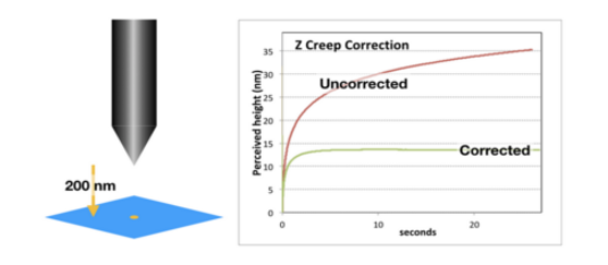 | ||
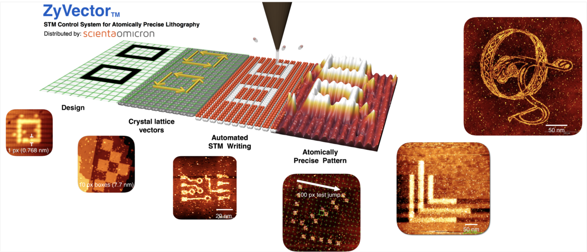
| Zyvex Lab’s ZyVector™ Control system provides the world’s highest (sub-nm) resolution lithography technology. Click here for more information | The Zyvex Creep and Hysteresis Correction Controller. Live tip position control for fast settling times after landing, and precise motion across the surface. Click here for more information. | ||
The 67th International Conference on Electron, Ion, and Photon Beam Technology and Nanofabrication
The 29th EIPBN Bizarre/Beautiful Micrograph Contest is now CLOSED. Please see all 2024 award winners and entries below.
The rules include the following:
- Entries have to be of a single image taken with a microscope and should not be significantly altered.
- There is no restriction with respect to the subject matter.
- Electron and ion micrographs have to be black and white.
In 2024, 46 entries were submitted from seven different countries.
The judges were:
- ShaChelle Manning – DARPA Chief Commericalization Strategist
- John Price – Price Farms Organics
- Anne Milasincic Andrews – UCLA
There were eight awards:
- Grand Prize
- Best Electron Micrograph
- Best Ion Micrograph
- Best Photon Micrograph
- Best Scanning Probe Micrograph
- Most Bizarre
- 3Beamers Choice
- Milligraph Award
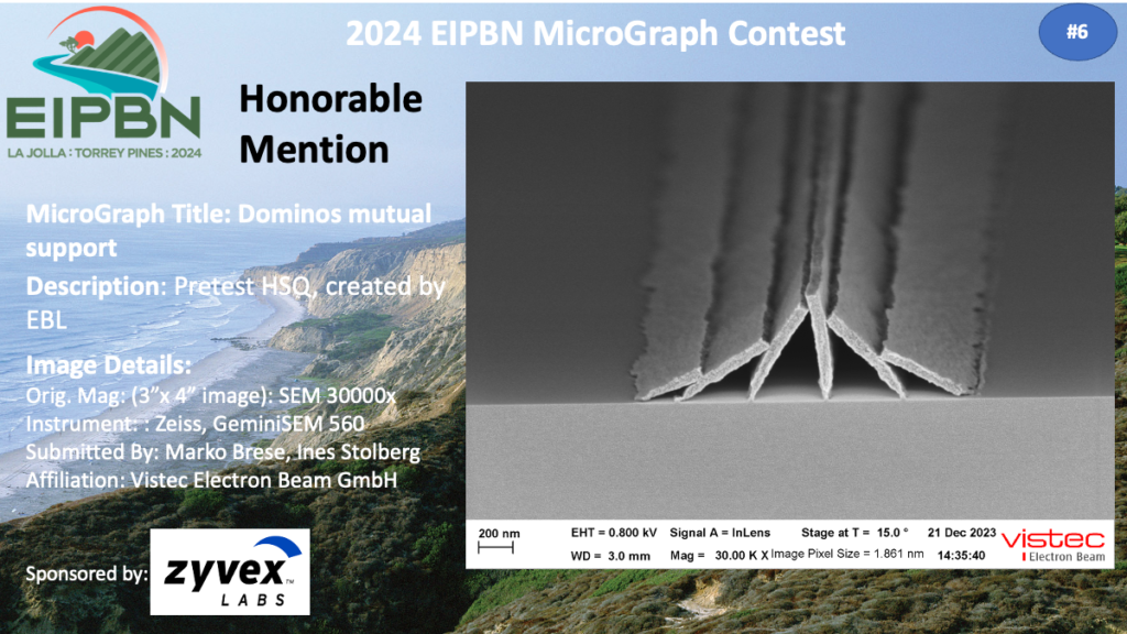
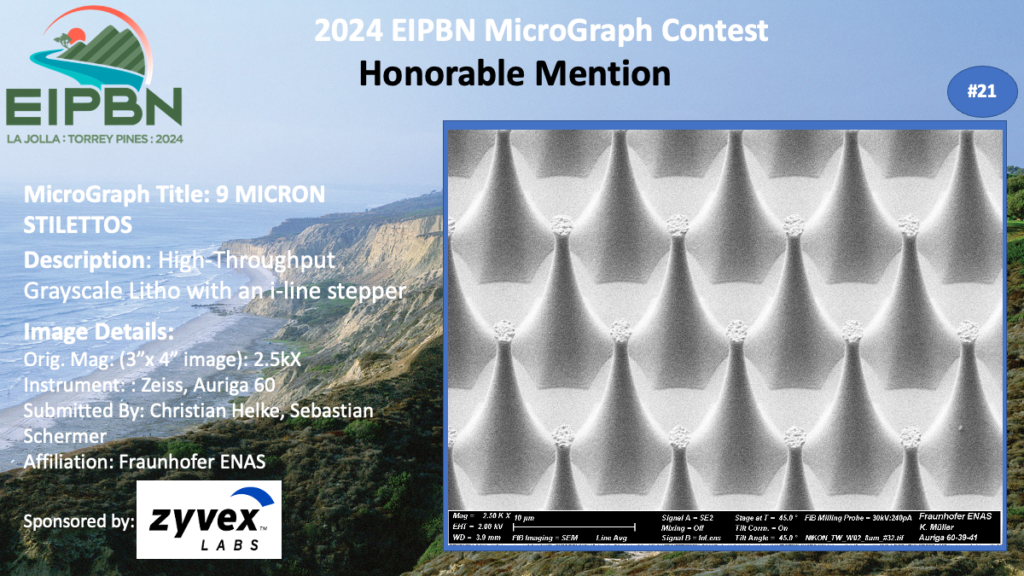
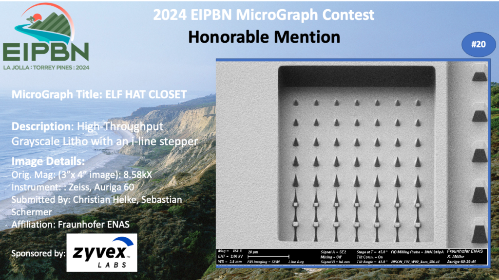
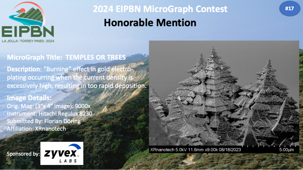
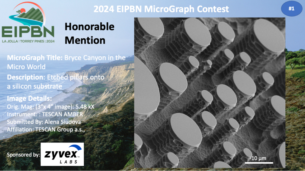
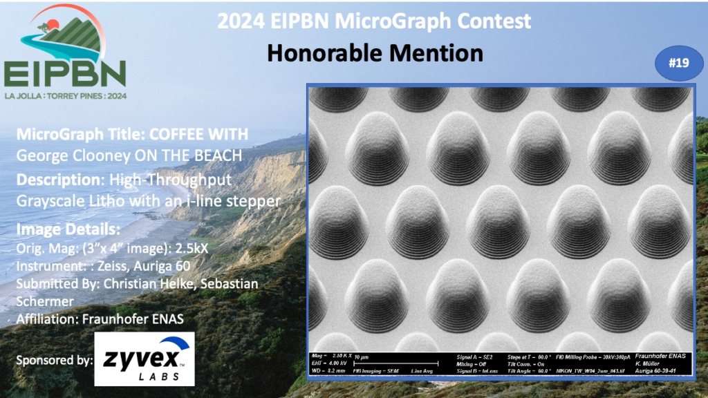
There were also six Honorable Mention awards given.
To download a PDF with all 46 entries, click below:
Grand Prize Micrograph
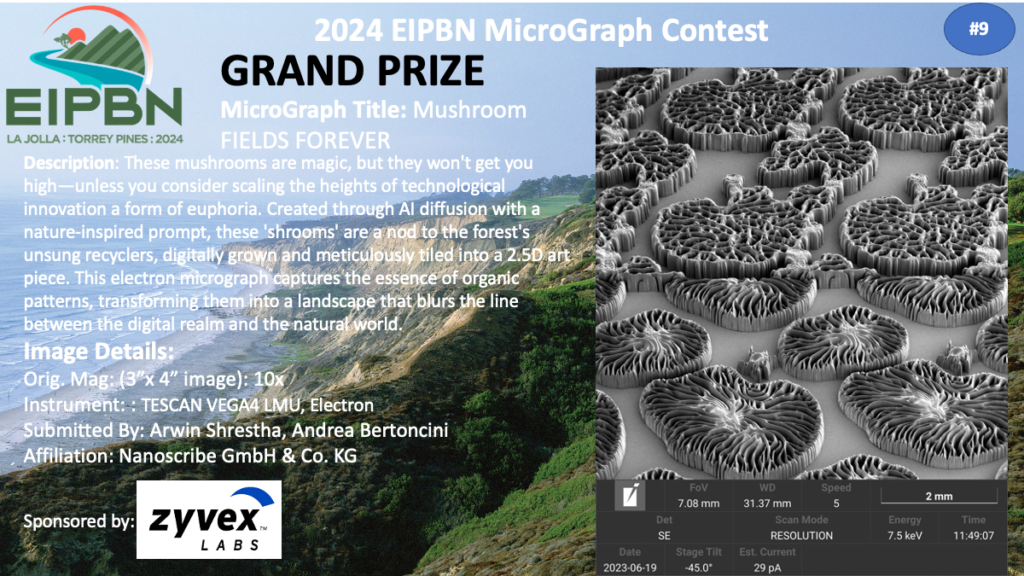
Title: Mushroom Fields Forever
Description:These mushrooms are magic, but they won’t get you high—unless you consider scaling the heights of technological innovation a form of euphoria. Created through AI diffusion with a nature-inspired prompt, these ‘shrooms’ are a nod to the forest’s unsung recyclers, digitally grown and meticulously tiled into a 2.5D art piece. This electron micrograph captures the essence of organic patterns, transforming them into a landscape that blurs the line between the digital realm and the natural world.
Magnification (3″ x 4″ image): 10X
Instrument: TESCAN VEGA4 LMU, Electron
Submitted by: Arwin Shrestha, Andrea Bertoncini
Affiliation: Nanoscribe GmbH & Co. KG
3-Beamers’ Choice & Best Electron Micrograph
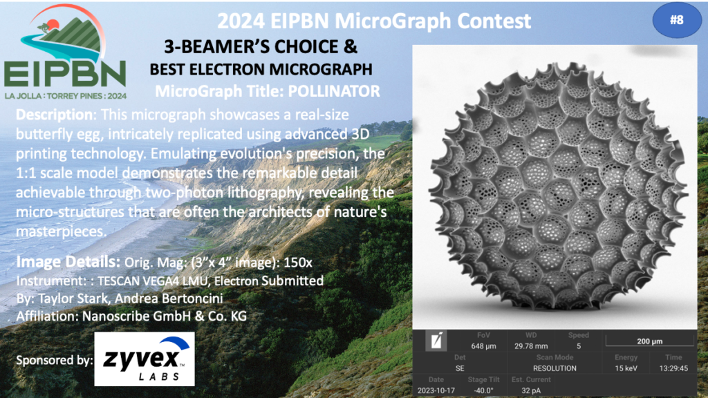
Title: Pollinator
Description: This micrograph showcases a real-size butterfly egg, intricately replicated using advanced 3D printing technology. Emulating evolution’s precision, the 1:1 scale model demonstrates the remarkable detail achievable through two-photon lithography, revealing the micro-structures that are often the architects of nature’s masterpieces.
Magnification (3″ x 4″ image): 150X
Instrument: TESCAN VEGA4 LMU, Electron
Submitted by: Taylor Stark, Andrea Bertoncini
Affiliation: Nanoscribe GmbH & Co. KG
Best Ion Micrograph
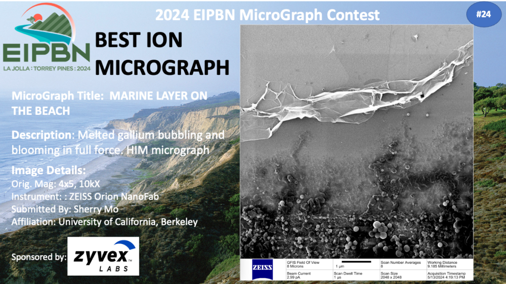
Title: Marine Layer on the beach
Description: Melted gallium bubbling and blooming in full force. HIM micrograph
Magnification (3″ x 4″ image): 10kX
Instrument: ZEISS Orion NanoFab
Submitted by: Sherry Mo,
Affiliation: University of California, Berkeley
Best Photon Micrograph
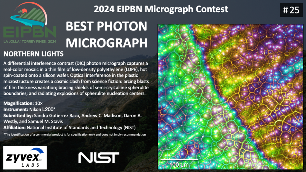
Title: Northern Lights
Description: A differential interference contrast (DIC) photon micrograph captures a real-color mosaic in a thin film of low-density polyethylene (LDPE), hot spin-coated onto a silicon wafer. Optical interference in the plastic microstructure creates a cosmic clash from science fiction: arcing blasts of film thickness variation; bracing shields of semi-crystalline spherulite boundaries; and radiating explosions of spherulite nucleation centers.
Magnification (3″ x 4″ image): 10X
Instrument: Nikon L200*
Submitted by: Sandra Gutierrez Razo, Andrew C. Madison, Daron A. Westly, and Samuel M. Stavis
Affiliation: National Institute of Standards and Technology (NIST)
Best Scanning Probe Micrograph
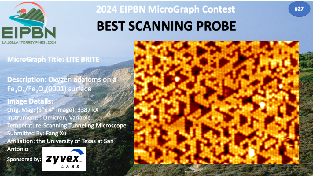
Title: Lite Brite
Description: Oxygen adatoms on a Fe3O4/Fe2O3(0001) surface
Magnification (3″ x 4″ image): 3387 kX
Instrument: ScientaOmicron VT STM
Submitted by: Fang Xu
Affiliation: University of Texas at San Antonio
Most Bizarre Micrograph
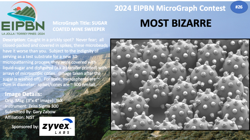
Title: Sugar Coated Mine Sweeper
Description: Caught in a prickly spot? Never fear; all closed-packed and covered in spikes, these microbeads have it worse than you. Subject to the indignity of serving as a test substrate for a new 3D micropatterning process, they were covered with liquid-sugar and disfigured (a.k.a transfer printed) with arrays of microscopic cones. (Image taken after the sugar is washed off). For scale, microspheres are ~ 7um in diameter; spikes/cones are ~ 900 nm tall.
Magnification (3″ x 4″ image): 3 kX
Instrument: Zeiss Sigma 300
Submitted by: Gary Zabow
Affiliation: NIST
Milligraph Award
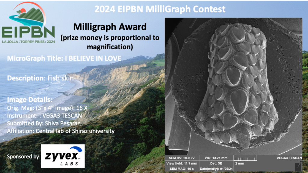
Title: I Believe in Love
Description: Fish Skin
Magnification (3″ x 4″ image): 16 X
Instrument: VEGA3 TESCAN
Submitted by: Shiva Pesaran
Affiliation: Central lab of Shiraz university
Honorable Mentions
