
←1998 |
2000→ |
The fields of research covered by this conference have been at the forefront of the drive to develop technology to make smaller and smaller structures. We have ventured into size regimes where we are often dependent on microscopes and the skill of microscopists to see the results of our work (and often what went wrong). To highlight the importance of micrographs to the field, the conference holds a micrograph contest. The entries were judged both from the technological and artistic standpoint. Six categories were defined:
- Best Electron Micrograph
- Best Ion Micrograph
- Best Photon Micrograph
- Best Scanning Probe MicroGraph
- Most Bizarre
- Grand Prize
In addition the judges selected one additional micrograph for an Honorable Mention Award.
The rules included the following:
- Contestants must have been registered 1999 conference attendees.
- Micrographs must be submitted as an 8 inch by 10 inch foil and must be accompanied by a completed entry sheet.
- Entries must be of a single image taken with a microscope and may not be significantly altered.
- There is no restriction with respect to the subject matter.
- Electron and ion micrographs must be black and white.
In 1999, 35 entries were submitted. There were many outstanding micrographs. The work represented in the submitted micrographs covered a wide range of fields including micro mechanical, photonic, and integrated circuit fabrication, chemical and dry etching, field emission tips, UV and x-ray optics, and of course e-beam, ion beam, x-ray, and photo lithography experiments. The panel of judges who selected the award winners consisted of:
Prof. Evelyn Hu
University of California at Santa Barbara
Nikki Marrion
World Bank
Dr. Kurt Ronse
Director of Lithography, IMEC, Belgium
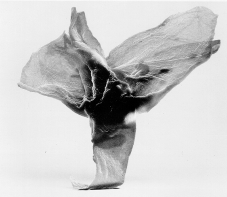
TITLE: Orchid
Description: Resist Particle – Shipley SPR 3625
Magnification for 3″x4″ image 1.8KX
Instrument: Hitachi 4100 SEM
Submitted by: Bobb Mohondro & Donna Whiteside Eaton SEO Fusion Systems Division
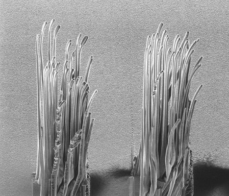
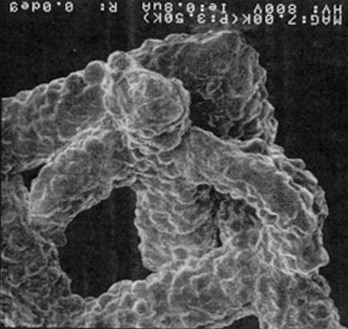
TITLE: Seagrass
Description: Attempts at etching copper did not work as planned.
Magnification for 3″x4″ image
Instrument: Micrion FIB
Submitted by: Nick Economou, Micrion
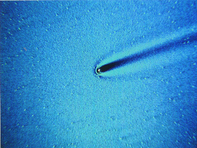
TITLE: Van Gogh in PMMA: Starry, Starry Night
Description: Optical photomicrograph of spin defects in PMMA
Magnification for 3″x4″ image: 62.5
Instrument: Leitz Ergolux 200 (darkfield image), Poloroid CCD still camera
Submitted by: Robert J. Davis Penn State EMPRL, University Park PA and Maggie A.Z. Hupcey Sabin Group, Bloomington, IN
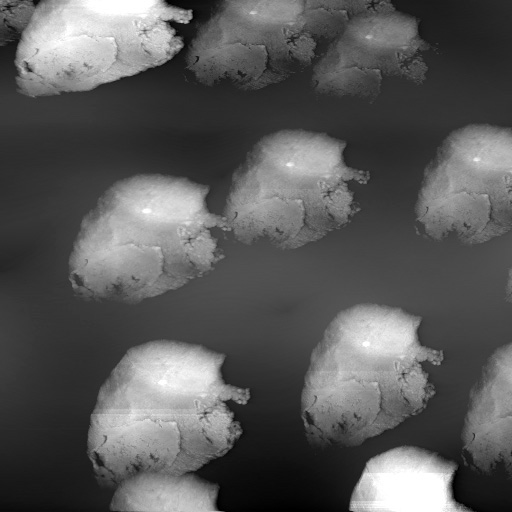
TITLE: Who’s imaging who here?
Description: Scanning tunneling microscope image of a silicon surface in which sample protrusions resulted in multiple tip images.
Magnification for 3″x4″ image: 127,000X
Instrument: Home made STM (Univ. of Maryland)
Submitted by: Andres Fernadez, ETEC Systems Inc.
TITLE: The Miracle Child
Description: First spotted in early 1991 on the tungsten grid of a Hitachi CD-SEM. The child lies peacefully with beckoning arms raised. Word of the miracle spread like wildfire throughout Yorktown Heights. Defective wafers were found to be healed simply by close proximity to the child.
Magnification for 3″x4″ image: 7KX
Instrument: Hitachi CD-SEM
Submitted by: Tim Brunner, IBM
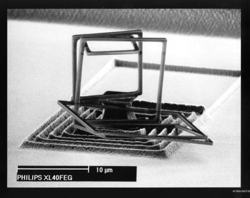
TITLE: Under Construction
Description: Under etching of the walls of this structure yielded this suprising construction.
Magnification for 3″x4″ image: 3000X
Instrument: Philips XL40 FEG SEM
Submitted by: J.P. Weterlings and F. Holthuysen, Philips
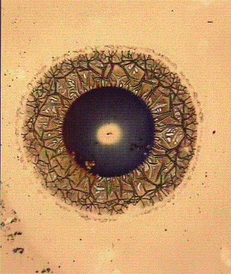
TITLE: Nano Eye
Description: A new approach to near-field optics.
Magnification for 3″x4″ image: 500X
Instrument: Leica Optical Microscope
Submitted by: Kathryn Wilder, Stanford University