
←2012 |
2014→ |
Bizarre/Beautiful
Micrograph Contest
“A good Micrograph is worth more than the MegaByte it consumes.”
Entries Presented by Dr. John Randall
The rules include the following:
• Entries have to be of a single image taken with a microscope and could not be significantly altered.
• There is no restriction with respect to the subject matter.
• Electron and ion micrographs have to be black and white.
In 2013, 70 entries were submitted.
The panel of judges who selected the award winners were:
• Don Tennant – Cornell
• Veronica Savu – University of Basel/EPFL
• Richard Blaikie – University of Canterbury
The Judges exercised their prerogative to liberally interpret the award categories, change the micrograph titles, and even rotate the micrograph if it pleased them.
There were six awards:
• Grand Prize
• Oops Micrograph
• Most Bizzare
• Best Ion Micrograph
• Best Electron Micrograph
• Most Entitled Micrograph
There were 11 Honorable Mentions.
All 2013 Entries (with original titles)
BEST ELECTRON MICROGRAPH
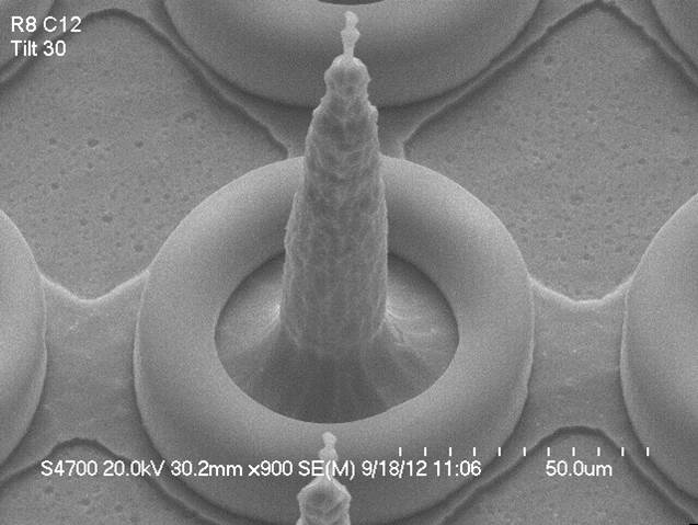
Title: The Tower of Bagel
Description: SEM image of tip of microelectrode with a Parylene C coating on it.
Magnification (3″x4″ image): 900X
Instrument (Make and Model): Hitachi S-4700 SEM
Submitted by: Bill Owen & J. Owen
Affiliation: Zyvex Labs
OOPS PRIZE
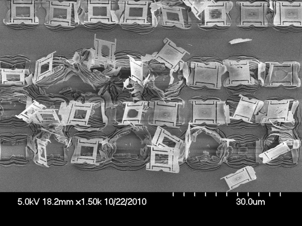
Title: University Yield Train Wreck
Description: GaP photonic devices with AlGaP sacrificial layer oxidized in air.
Magnification (3″x4″ image): 1.5 KX
Instrument: Hitachi 4700 SEM
Submitted by: Luozhou Li
Affiliation: Columbia University
BEST ION MICROGRAPH
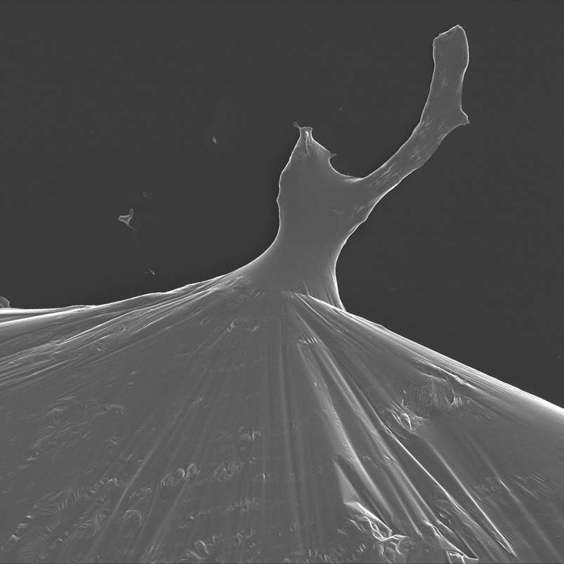
Title: Stickly Ballroom
Description: Helium ion image of dewetting of a liquid metal alloy with a thin oxide surface skin.
Magnification (4″ x 5″ image): 2.286 kX
Instrument: Carl Zeiss Orion
Submitted by: Kate Klein & Eva Mutunga
Affiliation: NIST / UDC
MOST BIZARRE MICROGRAPH
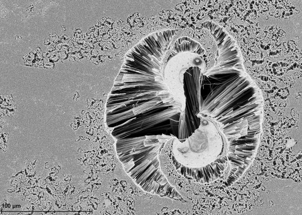
Title: Ceremonial Shrip Sex
Description: The strange top view of a carbon nanotube forest.
Magnification (3″x4″ image): 305X
Instrument: Zeiss Sigma VP
Submitted by: Mike Chang & Alireza Nojeh
Affiliation: Electrical and Computer Engineering, University of British Columbia
MOST ENTITLED MICROGRAPH
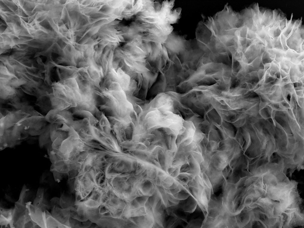
Title: Backdraft Where there’s smoke…Dave’s not here, French Can Can, Fur Balls, Cat on Fire with an Attitude.
Description: Re-crystalized AgNO3 salt and SDS surfactant combine to create flowing structures resembling flower petals. Silver salts and surfactants are used for in-situ liquid phase deposition studies in an ESEM. After experiments are completed, unconsumed reactants are washed away; however, some reactants tend to remain and re-crystalize. Many of the re-crystalized structures have straight edges, whiel this unique structure displays intricate curvature.
Magnification (3″x4″ image): 2.8 KX
Instrument: FEI Quanta FEG
Submitted by: Matthew Bresin
Affiliation: University of Kentucky
GRAND PRIZE
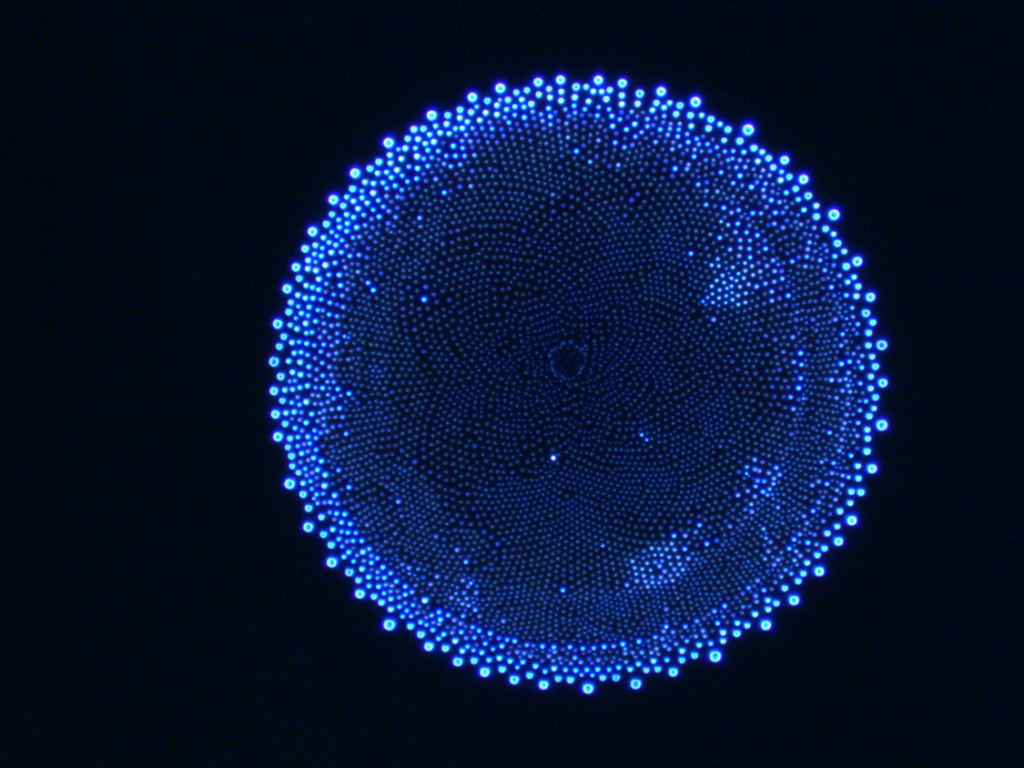
Title: Blue Sun Flower (-90K)
Description: Field image of residue from HSQ, water, and TMAH on silicon substrate.
Magnification (3″x4″ image):10X
Instrument: Olympus MX61
Submitted by: Devin K. Brown
Affiliation: Georgia Tech
HONORABLE MENTION
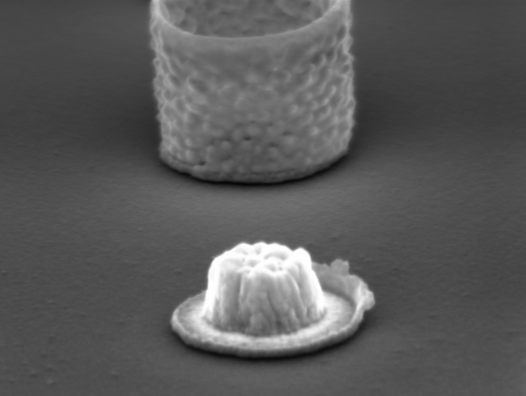
Title: Nexpresso, what else?
Description: By an innovative process combining nanoimprint and reactive ion etching, we were able to fabricate this kind of gold nanocups filled with aluminum (structure on the back). One of the nanocups lost its walls, remaining only the base of the cup and the inside’s aluminum (front capsule).
Magnification (3″x4″ image): 250 kX
Instrument: Zeiss Auriga
Submitted by: Nerea Alayo
Affiliation: IMB-CNM (CSIC)
HONORABLE MENTION
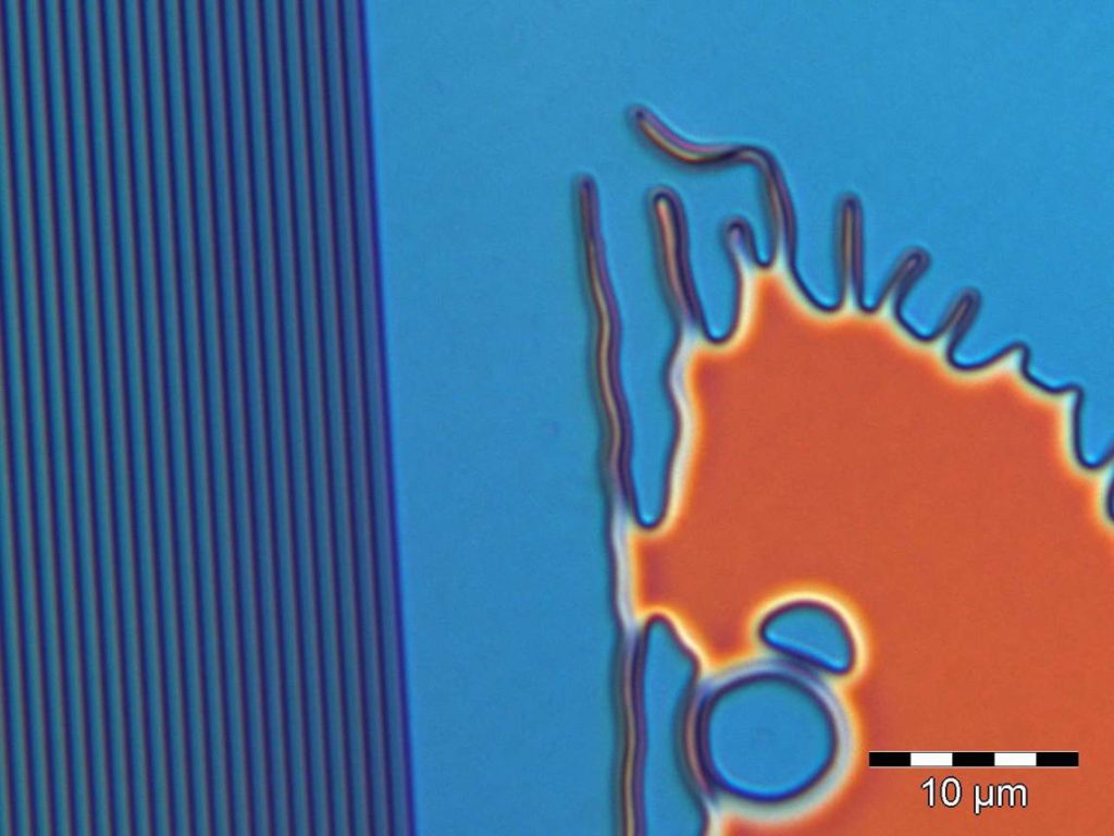
Title: Micro Seahorse
Description: Impact of intensity swing curves within photoresist layers.
Magnification (3″x4″ image): 700X
Instrument: Optical Microscope
Submitted by: Dhima Khalid
Affiliation: University of Wuppertal
HONORABLE MENTION
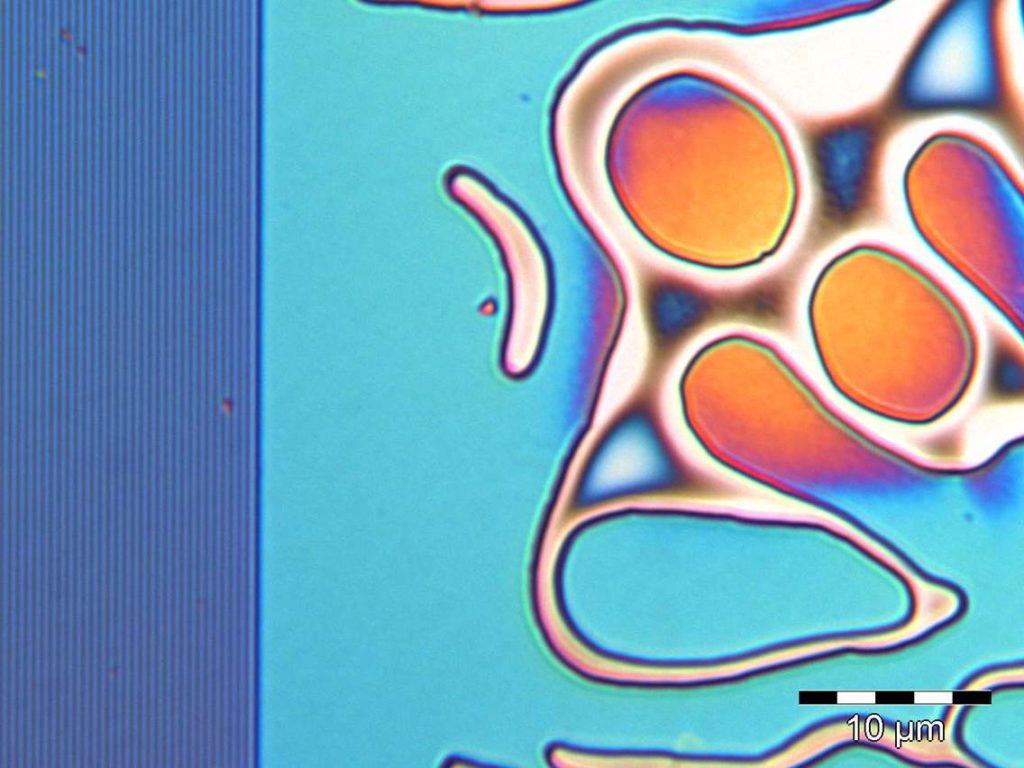
Title: Ahh!! The Boomerang does come back!
Description: Impact of intensity swing curves within photoresist layers.
Magnification (3″x4″ image): 700X
Instrument: Optical Microscope
Submitted by: Dhima Khalid
Affiliation: University of Wuppertal
HONORABLE MENTION
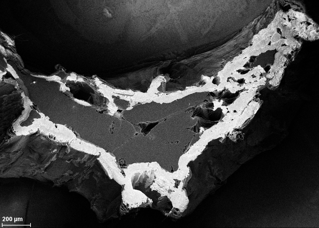
Title: Honshu of Japan
Description: Caesium-encapsulated carbon nanotube film, prepared by immersing in cesium carbonate solution.
Magnification (3″x4″ image): 46 X
Instrument (Make and Model): Zeiss Sigma VP
Submitted by: Mike Chang & Alireza Nojeh
Affiliation: Electrical and Computer Engineering, University of British Columbia
HONORABLE MENTION
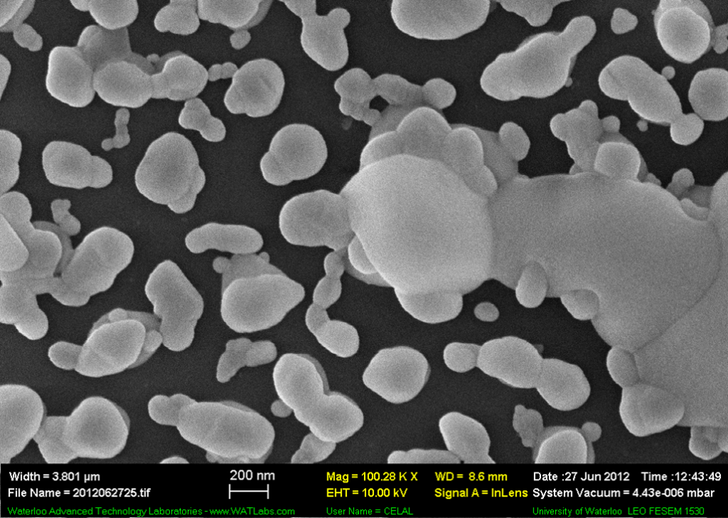
Title: Yummy Yummy!
Description: SEM image of self-asembled CsCl film evaporated by thermal evaporation technique.
Magnification (3″x4″ image): 100.28 kX
Instrument (Make and Model): Zeiss LEO 1530 Gemini
Submitted by: Celal Con
Affiliation: University of Waterloo
HONORABLE MENTION
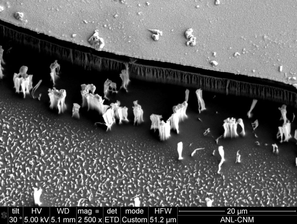
Title: Brighton Beach
Description: SEM MicroGraph of polymer etched via aluminum oxide membrane.
Magnification (3″x4″ image): 2700 X
Instrument (Make and Model): FEI Nova 600 SEM
Submitted by: Olga V. Makarova & Ralu Divan
Affiliation: Creatv Microtech Inc., Argonne National Laboratory
HONORABLE MENTION
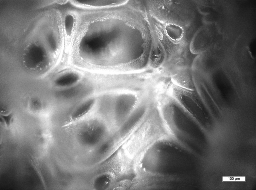
Title: SCREAM
Description: Image taken during bubble and pore size count in research to correlate whipping power and the foaming of egg white (dried in this micrograph).
Magnification (3″x4″ image): Unknown
Instrument (Make and Model): Leica DM4000M
Submitted by: Ka C. Wong
Affiliation: Evans Analytical Group
HONORABLE MENTION
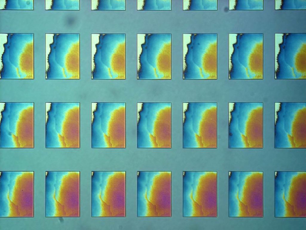
Title: Watching Hair Grow
Description: A test of field illumination uniformity oer a range of dose values, in a thin resist layer.
Magnification (3″x4″ image): 40 X
Instrument (Make and Model): Nikon L200
Submitted by: Steven Hickman
Affiliation: Harvard CNS
HONORABLE MENTION
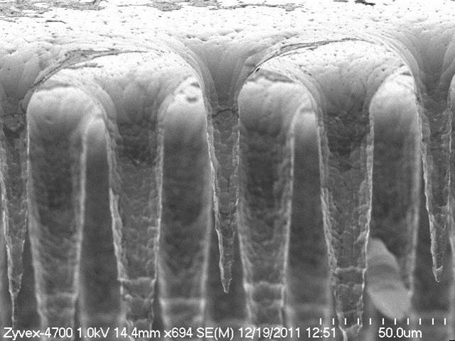
Title: Stalactites
Description: SEM image of Deep Reactive Ion Etched (DRIE) microelectrodes fabricated in SCS.
Magnification (3″x4″ image): 694 X
Instrument (Make and Model): Hitachi S-4700 SEM
Submitted by: J. Owen & Justin Alexander
Affiliation: Zyvex Labs
HONORABLE MENTION
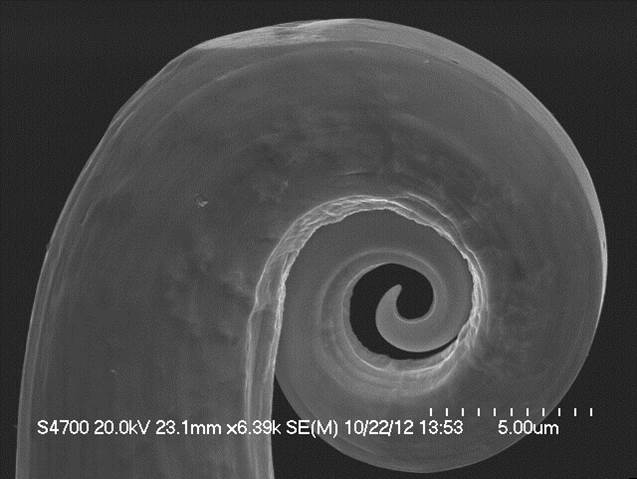
Title: Koru
Description: SEM image of a polycrystalline STM tip that has been crashed and curled.
Magnification (3″x4″ image): 6.39 kX
Instrument (Make and Model): Hitachi S-4700 SEM
Submitted by: Bill Owen & J. Owen
Affiliation: Zyvex Labs
HONORABLE MENTION
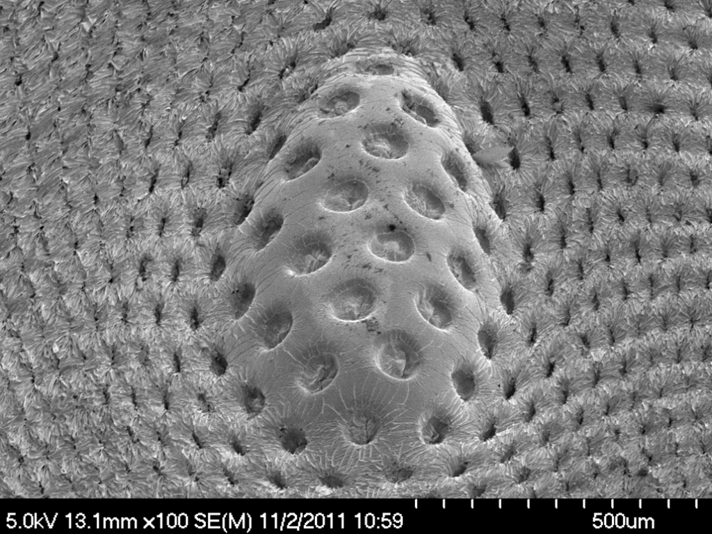
Title: Hemorrhoid
Description: An artificial compound eyes using novel microfabrication method of polymer.
Magnification (3″x4″ image): 100 X
Instrument (Make and Model): Hitachi SEM 4700
Submitted by: Huan Hu
Affiliation: Electrical & Computer Dept., University at Urbana-Champaign